
Características clinicopatológicas de nódulos pulmonares: Experiencia en Clínica Reina Sofía, Bogotá, Colombia
DOI:
https://doi.org/10.30944/20117582.903Palabras clave:
neoplasias pulmonares, nódulo pulmonar solitario, biopsia con aguja, patología, diagnóstico, diagnóstico por imagenResumen
Introducción. El cáncer de pulmón es la primera causa de mortalidad por cáncer a nivel mundial, lo que hace que sea considerado un problema de salud pública. Existen diferentes hallazgos imagenológicos que hacen sospechar la presencia de cáncer de pulmón, uno de los cuales son los nódulos pulmonares; sin embargo, estos también pueden verse en entidades benignas.
Métodos. Se incluyeron 66 pacientes con biopsia de nódulo pulmonar en la Clínica Reina Sofía, en la ciudad de Bogotá, D.C., Colombia, entre el 1° de marzo del 2017 y el 28 de febrero del 2020. Se analizaron las características demográficas de los pacientes, las características morfológicas e histopatológicas de los nódulos pulmonares y la correlación entre sus características imagenológicas e histopatológicas.
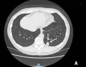
Resultados. El 69,2 % de los nódulos estudiados tenían etiología maligna, de estos el 55,5 % era de origen metástasico y el 44,5 % eran neoplasias primarias de pulmón, con patrón sólido en el 70,6 % de los casos. El patrón histológico más frecuente fue adenocarcinoma. Respecto a las características radiológicas, en su mayoría los nódulos malignos medían de 1 a 2 cm, de morfología lisa y distribución múltiple, localizados en lóbulos superiores.
Conclusiones. La caracterización de los nódulos pulmonares brinda información relevante que orienta sobre los diagnósticos más frecuentes en nuestro medio, cuando se estudian nódulos sospechosos encontrados incidentalmente o en el seguimiento de otro tumor. Como el nódulo es la manifestación del cáncer temprano del pulmón, establecer programas de tamización que permitan el diagnóstico oportuno, es hoy día una imperiosa necesidad, para reducir la mortalidad.
Descargas
Referencias bibliográficas
Siegel R, Naishadham D, Jemal A. Cancer statistics, 2012. CA Cancer J Clin. 2012;62:10-29. https://doi.org/10.3322/caac.20138
World Health Organization. International Agency for Research on Cancer. Cancer today. Fecha de consulta: 18 de febrero de 2021. Disponible en: https://gco.iarc.fr/today/home
Ministerio de Salud y Protección Social. Guía de práctica clínica para la detección temprana, diagnóstico, estadificación y tratamiento del cáncer de pulmón. Guía para profesionales de la salud. Bogotá D.C; 2014 p. 2014 - Guía No. 36. Fecha de consulta: 18 de Enero de 2021. Disponible en: https://www.sispro.gov.co/ observatorios/oncancer/Paginas/publicaciones-interes-piel-no-melanoma.aspx
Bach PB, Mirkin JN, Oliver TK, Azzoli CG, Berry DA, Brawley OW, et al. Benefits and harms of CT screening for lung cancer. JAMA. 2012;307:2418-29. https://doi.org/10.1001/jama.2012.5521
Sihoe ADL, Cardillo G. Solitary pulmonary ground-glass opacity: is it time for new surgical guidelines? European Journal of Cardio-Thoracic Surgery. 2017;52:848-51. https://doi.org/10.1093/ejcts/ezx211
The National Lung Screening Trial Research Team. Reduced lung-cancer mortality with low-dose computed tomographic screening. N Engl J Med. 2011;365:395- 409. https://doi.org/10.1056/NEJMoa1102873
Bray F, Ferlay J, Soerjomataram I, Siegel RL, Torre LA, Jemal A. Global cancer statistics 2018: GLOBOCAN estimates of incidence and mortality worldwide for 36 cancers in 185 countries. CA Cancer J Clin. 2018;68:394- 424. https://doi.org/10.3322/caac.21492
Houghton AM. Mechanistic links between COPD and lung cancer. Nat Rev Cancer. 2013;13:233-45. https://doi.org/10.1038/nrc3477
Hammer MM, Hatabu H. Subsolid pulmonary nodules: Controversy and perspective. Eur J Radiol Open. 2020;7:100267. https://doi.org/10.1016/j.ejro.2020.100267
Wang W, Xie M, Dou S, Cui L, Zheng CY, Xiao W. The link between chronic obstructive pulmonary disease phenotypes and histological subtypes of lung cancer: a case - control study. International Journal of COPD. 2018;13:1167-75. https://doi.org/10.2147/COPD.S158818
MacMahon H, Naidich DP, Goo JM, Lee KS, Leung ANC, Mayo JR, et al. Guidelines for management of incidental pulmonary nodules detected on CT images: From the Fleischner Society 2017. Radiology. 2017;284:228-43. https://doi.org/10.1148/radiol.2017161659
Chen D, Dai C, Kadeer X, Mao R, Chen Y, Chen C. New horizons in surgical treatment of ground-glass nodules of the lung: experience and controversies. Ther Clin Risk Manag. 2018;14:203-11. https://doi.org/10.2147/TCRM.S152127
Takasugi J, Godwin J. The Solitary Pulmonary Nodule: Radiologic Assessment. In: Sperber M (eds). Radiologic Diagnosis of Chest Disease. Springer, London. https://doi.org/10.1007/978-1-4471-0693-7_32
McWilliams A, Tammemagi M, Mayo J, Roberts H, Liu G, Soghrati K et al. Probability of Cancer in Pulmonary Nodules Detected on First Screening CT. N Engl J Med. 2013;369:910-9. https://doi.org/10.1056/NEJMoa1214726
Gould MK, Donington J, Lynch WR, Mazzone PJ, Midthun DE, Naidich DP, Wiener RS. Evaluation of individuals with pulmonary nodules: when is it lung cancer? Diagnosis and management of lung cancer, 3rd ed: American College of Chest Physicians evidence-based clinical practice guidelines. Chest. 2013;143:e93S-e120S. https://doi.org/10.1378/chest.12-2351
Seo JB, Im JG, Goo JM, Chung MJ, Kim MY. Atypical pulmonary metastases: Spectrum of radiologic findings. Radiographics. 2001;21:403-17. https://doi.org/10.1148/radiographics.21.2.g01mr17403
Ost D, Fein AM, Feinsilver SH. The Solitary Pulmonary Nodule. N Engl J Med. 2003;348:2535-42. https://doi.org/10.1056/NEJMcp012290
Barrio JL, Suarez M, Rodriguez JL, Saldana MJ, Pitchenik AE. Pneumocystis carinii pneumonia presenting as cavitating and noncavitating solitary pulmonary nodules in patients with the acquired immunodeficiency syndrome. Am Rev Respir Dis. 1986;134:1094-6. https://doi.org/10.1164/arrd.1986.134.5.1094
Echeverri A, Long RF, Check W, Burnett CM. Pulmonary dirofilariasis. The Ann Thorac Surg. 1999;67:201-2. https://doi.org/10.1016/s0003-4975(98)01060-1
Guo W, Zhao YP, Jiang YG, Wang RW, Ma Z. Surgical treatment and outcome of pulmonary hamartoma: a retrospective study of 20-year experience. Journal of Experimental & Clinical Cancer Research. 2008;27:8. https://doi.org/10.1186/1756-9966-27-8
Dai J, Yu G, Yu J. Can CT imaging features of groundglass opacity predict invasiveness? A meta-analysis. Thoracic Cancer. 2018;9:452-8. https://doi.org/10.1111/1759-7714.12604
Maj GT, Gracey DR, Byrd RB. The management and evaluation of the solitary pulmonary nodule. Chest. 1974;66:236-9. https://doi.org/10.1378/chest.66.3.236
Gruden JF, Ouanounou S, Tigges S, Norris SD, Klausner TS. Incremental benefit of maximum-intensity-projection images on observer detection of small pulmonary nodules revealed by multidetector CT. Am J Roentgenol. 2002;179:149-57. https://doi.org/10.2214/ajr.179.1.1790149
Gould MK, Ananth L, Barnett PG. A clinical model to estimate the pretest probability of lung cancer in patients with solitary pulmonary nodules. Chest. 2007;131:383-8. https://doi.org/10.1378/chest.06-1261
Gohagan J, Marcus P, Fagerstrom R, Pinsky P, Kramer B, Prorok P, for The Lung Screening Study Research Group. Baseline findings of a randomized feasibility trial of lung cancer screening with spiral CT scan vs chest radiograph. The Lung Screening Study of the National Cancer Institute. Chest. 2004;126:114-21. https://doi.org/10.1378/chest.126.1.114
Gurney JW. Determining the likelihood of malignancy in solitary pulmonary nodules with Bayesian analysis. Part I. Theory. Radiology. 1993;186:405-13. https://doi.org/10.1148/radiology.186.2.8421743
Lee JE, Jeong WG, Kim YH. Differentiation of primary lung cancer from solitary lung metastasis in patients with colorectal cancer: a retrospective cohort study. World Journal of Surgical Oncology. 2021;19:28. https://doi.org/10.1186/s12957-021-02131-7
Vega J, Lazo D, Undurraga F, Clavero JM, Rodríguez P. Caracterización de nódulos pulmonares resecados. Experiencia de manejo por un programa multidisciplinario. Rev méd Chile. 2018;146:1261-8. http://dx.doi.org/10.4067/S0034-98872018001101261
Snoeckx A, Reyntiens P, Desbuquoit D, Spinhoven MJ, Van Schil PE, van Meerbeeck JP, Parizel PM. Evaluation of the solitary pulmonary nodule: size matters, but do not ignore the power of morphology. Insights into Imaging. 2018;9:73-86. http://doi.org/10.1007/s13244-017-0581-2
Ost D, Fein A. Management strategies for the solitary pulmonary nodule. Curr Opin Pulm Med. 2004;10:272- 8. http://doi.org/10.1097/01.mcp.0000130322.11513.c8
Oke JL, Pickup LC, Declerck J, Callister ME, Baldwin D, Gustafson J, et al. Development and validation of clinical prediction models to risk stratify patients presenting with small pulmonary nodules: a research protocol. Diagn Progn Res. 2018;2:22. https://doi.org/10.1186/s41512-018-0044-3
Zhang M, Kono M. Solitary pulmonary nodules: evaluation of blood flow patterns with dynamic CT. Radiology. 1997;205:471-8. https://doi.org/10.1148/radiology.205.2.9356631
Kiranantawat N, McDermott S, Fintelmann FJ, Montesi SB, Price MC, Digumarthy SR, Sharma A. Clinical role, safety and diagnostic accuracy of percutaneous transthoracic needle biopsy in the evaluation of pulmonary consolidation. Respir Res. 2019;20:23. https://doi.org/10.1186/s12931-019-0982-5
Clark ME, Young B, Bedford LE, das Nair R, Robertson JFR, Vedhara K, et al. Lung cancer screening: does pulmonary nodule detection affect a range of smoking behaviours? J Public Health (Oxf). 2018;41:600-8. https://doi.org/10.1093/pubmed/fdy158
Descargas
Publicado
Cómo citar
Número
Sección
Licencia
Derechos de autor 2021 Revista Colombiana de Cirugía

Esta obra está bajo una licencia internacional Creative Commons Atribución-NoComercial-SinDerivadas 4.0.
Todos los textos incluidos en la Revista Colombiana de Cirugía están protegidos por derechos de autor. Las opiniones expresadas en los artículos firmados son las de los autores y no coinciden necesariamente con las de los directores o los editores de la Revista Colombiana de Cirugía. Las sugerencias diagnósticas o terapéuticas como elección de productos, dosificación y métodos de empleo corresponden a la experiencia y al criterio de los autores.


.jpg)